QUT researchers are leading the world in new mathematical experiments to better understand changes in melanomas in real-time.
The findings of their new research have been published in the UK-based .
The researchers developed mathematical models never created before by studying lab-based populations of cancer cells using fluorescent technology and simulating growth patterns.
Cases of melanoma are reported to increase by .
In Queensland and New Zealand has surpassed Australia with 50 cases per 100,000 people, according to the Cancer Council.
QUT’s works at the intersection of maths and biology and led the team of researchers from Queensland and New Zealand.
The study is aimed at quickly identifying key features of lab-grown tumour growth, known as tumour spheroids, to provide potential treatments as opposed to waiting days for the cells to do their own thing.
- Lab-grown populations of cells, known as spheroids, mimic how a tumour behaves, mimicking the availability of nutrients and waste within the population as cells grow, age, live, and die.
- Modelling shows changes with the size of the spheroid.
- Modelling enables better decisions about experiment design by simulating what happens to these spheroids very quickly.
- Makes experiments ‘reproducible’ because we know exactly what the mathematical model does.
“The mathematical models had not been developed before this research as the biotechnology is new,” Professor Simpson said.
Professor Simpson said by using real-time fluorescent cell cycle labelling technology, we can track the life cycle, or age, of individual cells within a complicated environment, like a tumour.
“We use computer simulations to study the role of tumour size and cell life cycle stage, and we simulate every single cell in the experimental tumour, carefully labelling it in the simulations so it can be matched with the appropriate fluorescent colour,” he said.
“Just after cell division, young cells fluoresce red and as cells progress through the cell cycle older cells fluoresce green. So, when you look at a population of melanoma cells in a tumour, we can see new information.
“We can see whether cells at the edge of the tumour are dividing, or whether cell division takes place deeper within the tumour.”

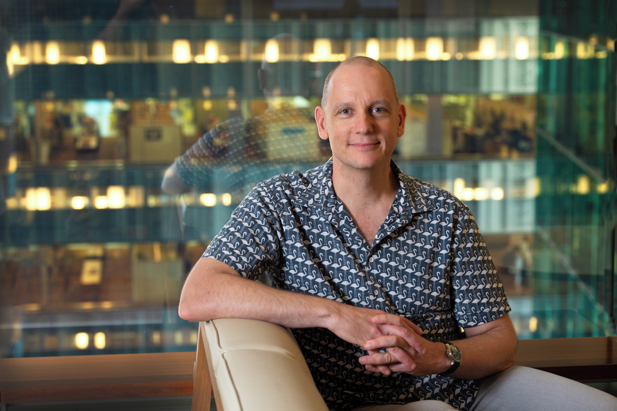
Professor Simpson (pictured above) said attacking biological problems using a mathematical and quantitative approach is useful for experimental scientists as it provided a ‘bigger picture’ by enabling us to rapidly explore the effects of population size, experimental duration, or nutrient availability.
“Cancer drugs only cover one part of melanoma treatments which is only one part of the story,” he said.
“By revolutionising the way, we track cancer cells within a realistic population in real time using mathematical modelling we obtain deeper understanding of basic cell biology processes, which can inform how experiments are performed and how experiments are interpreted.”
The investigation was prompted through an ARC Discovery grant Professor Simpson was awarded in 2020. He is a co-leader of the Models and Algorithms program in .
The journal articles co-authors include Jonah Klowss, Alexander Browning, Ryan Murphy and Elliot Carr from , Michael Plank from University of Canterbury, Te Punaha Matatini from New Zealand Centre of Research Excellence in Complex Systems and Data Analytics and Gency Gunasingh and Nikolas Haass from UQ’s Diamantina Institute.
A pdf of the journal article is available upon request.
MEDIA RESOURCES: Images, including an animation of the tumour spheroid, is available to download .


