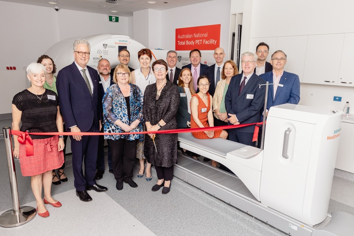
The officially opens in Sydney today, delivering the first Total Body Positron Emission Tomography (TB-PET) scanner for Australia-wide open access research, as well as clinical use.
The facility will drive advancements in cancer studies, neurological disorders, cardiovascular disease and drug development, and reduce scanning time and radiation doses to transform patient care.
The $15 million facility is a collaborative venture between the University of Sydney, the (NIF) and , to benefit Australian patients, clinicians, researchers and industry partners.
Located at Royal North Shore Hospital, the Siemens Biograph Vision Quadra is a revolutionary leap forward in nuclear medical imaging. This cutting-edge device enables comprehensive whole-body imaging in a single scan, significantly reducing radiation exposure and cutting down scanning time from 20 minutes to as little as three, all while delivering higher-quality images.
The ability to scan all tissues and organs simultaneously offers unique insights into whole-body physiology and interactions between organs that no other clinical imaging technology can provide. It presents research opportunities across a wide range of medical applications, such as oncology, neuroscience, cardiology, infectious diseases, and drug discovery – including exploring complex human biology and the way multiple organs interact such as the brain-gut axis.
The facility is Australia’s most sensitive PET scanner dedicated to research, and will be a critical tool for clinical trials and industry collaborations. The ability to image the entire human body allows researchers to observe drug absorption, accumulation and elimination processes in all organs simultaneously.
Reduced radiation and scanning times expand PET imaging options for vulnerable groups such as children in impactful research and clinical studies. It also encourages the participation of healthy individuals in clinical trials and enables repeated scanning of patients to better understand disease progression and treatment effects, broadening medical research insights.
“The collaboration between the University of Sydney, the ³Ô¹ÏÍøÕ¾ Imaging Facility and Northern Sydney Local Health District demonstrates the power of partnerships in driving innovation,” said Professor Mark Scott, Vice-Chancellor and President of the University of Sydney.
“This facility shows what can be achieved when leading institutions join forces to advance healthcare and research capabilities. We are not only improving the health of patients today, but also utilising this technology to fast-track new discoveries for the future.”
Australian ³Ô¹ÏÍøÕ¾ Total Body PET Facility
A game changer in medical imaging.
One such study is examining how the molecule oxytocin impacts the brain and body when delivered to humans. Oxytocin is one of the most important natural chemicals in the brain that guides social behaviour. When administered, research shows it can improve social understanding and may have benefits to support people with schizophrenia and autism. However, it is a mystery about where oxytocin is absorbed and the circuits it impacts in the brain and body to cause its effects in humans.
Using the TB PET Scanner, a team led by the University of Sydney’s Professor Adam Guastella, will see in real-time the brain and body circuits impacted by oxytocin after its delivery intranasally or by intravenous injection. This has the potential to change fundamental knowledge of the biology of human social behaviour and could lead to a range of new therapies.
The new facility forms part of the University of Sydney’s Core Research Facility for biomedical imaging. As a nationally significant research platform, it is also a flagship of the ³Ô¹ÏÍøÕ¾ Imaging Facility (NIF), through the Australian Government Department of Education’s ³Ô¹ÏÍøÕ¾ Collaborative Research Infrastructure Strategy (NCRIS).
Governing Board Chair of the ³Ô¹ÏÍøÕ¾ Imaging Facility, Professor Margaret Harding, said the NIF’s investment of $8m in the Australian ³Ô¹ÏÍøÕ¾ Total Body PET Facility was its largest to date, and represented Australia’s largest single investment in molecular imaging, underpinning research that is of high priority in reducing Australia’s burden of disease.
“The facility is a unique national asset which will revolutionise Australia’s capacity to attract and support research and industry undertaking clinical trials for the development of new pharmaceuticals and medical products to improve health outcomes for Australia,” Professor Harding said.
The Australian ³Ô¹ÏÍøÕ¾ Total Body PET Facility will operate under an equal time-share arrangement between clinical use and research, ensuring five day per week open access for all researchers throughout Australia.
Speaking to patient benefits, Chief Executive of the Northern Sydney Local Health District, Adjunct Professor Anthony Schembri, said: “Royal North Shore Hospital and Northern Sydney Local Health District have a proud history of delivering world-class imaging and care to improve patient outcomes.
“We are extremely honoured to be hosting this Australian-first where patients can receive world class care, and researchers can use the scanner for clinical research which may translate into improving patient care in the future.”
The University’s contribution to the new facility is underpinned by a bequest made by William Chapman who left the majority of his estate as a gift dedicated to cancer research at the University of Sydney. His legacy is set to have an enormous impact on cancer research and on the survival and quality of life of patients.
The University of Sydney’s Professor Emma Johnston, Deputy Vice-Chancellor (Research), said: “The combined clinical and research arrangements for this amazing medical imaging technology and its location in a bustling hub of activity at Royal North Shore Hospital will foster collaboration among researchers, healthcare providers, policymakers, and industry leaders to fast-track innovation in research translation.”


