Breakthrough microbeam radiation therapy technique draws a bead on hard to treat tumours

A new radiation therapy technique pioneered by scientists from the University of Wollongong’s (CMRP) has shown promise for improving treatment outcomes in patients with brain cancer.
Working at the Australian Synchrotron facility in Melbourne, the scientists tested a technique for the treatment of high-grade brain cancer using personalised microbeam radiation therapy (MRT), combining it with an innovative assessment of tumour dose-coverage.
MRT uses ultra-fine X-rays – each smaller in diameter than a human hair – to destroying the cancerous tissue while not harming the surrounding healthy tissue. Precise targeting also enables much higher dosages to be delivered to the tumour in a very short time.
The researchers used CT scans, performed at Monash Biomedical Imaging, to map individual brain tumours in rats, and then used MRT to deliver high dose to the cancer cells with pinpoint precision. The synchrotron is able to produce much more powerful X-rays than conventional hospital X-ray machines.
The MRT treated rats survived for significantly longer than non-irradiated rats with the same aggressive brain tumours. No long-term adverse effects were observed following MRT, and there was no noticeable decline in cognition, vision, mobility, or behaviour in the treated rats.
The , which included researchers from the (IHMRI), Australian Synchrotron – Australia’s Nuclear Science and Technology Organisation (ANSTO), Central Coast Cancer Centre and Prince of Wales Hospital, is published in Scientific Reports.
It is the first long-term Australian MRT brain cancer survival study, and the first in the world to look at optimisation of personalised pre-clinical MRT of high-grade brain cancer. The results and methods investigated MRT from multiple points of view including radiation and medical physics, radiobiology, diagnostic imaging, and preclinical survival.
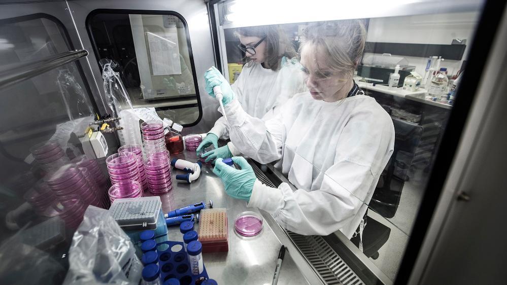
Lead author and UOW PhD student Elette Engels (pictured above) said brain tumours were among the most difficult cancers to treat.
“Brain cancers require more rigorous and novel treatment strategies to overcome their radiation resistance,” she said.
“This new MRT technique treats tumours with very narrow wafer-like X-ray blades to deliver very high doses of synchrotron radiation delivered in a very short time.
“This is not feasible with conventional radiotherapy X-ray machines in hospitals. Our research shows that the treatment of tumour cells is much more effective when the radiation dose is delivered using MRT.
“Our work aims to optimise this technique and personalise the entire procedure, from diagnosis to treatment, for each patient.”
Treating brain cancers in children and young adults is especially difficult. Over the past 30 years, treatment outcomes for brain cancer in children and young adults have remained at a stand-still.
Corresponding author Dr said that despite advances in surgical techniques, radiotherapy and chemotherapeutics, brain tumours remain difficult to remove surgically and can be resistant to radiation and drug treatments.
“A breakthrough in the treatment of brain cancer is well overdue,” Dr Tehei said.
“Many brain cancer survivors suffer from cognitive and somatic side effects of the treatment, with increased risks in children.
“Sparing normal tissue from damage is key to improved quality of life for brain cancer survivors.”
Personalised synchrotron MRT holds the promise of quicker, more effective treatment of brain cancers.
Current radiation therapy for a brain tumour is typically delivered over several weeks with daily radiation treatments. Instead of hitting a larger area of the brain with lower doses of X-rays, repeated numerous times, the new technique would involve a single dose of ultra high dose rate X-rays, precisely targeted at the cancerous cells.
“A single dose of this personalised synchrotron MRT treatment could be more effective than multiple radiation treatments as they are delivered now. Waiting times and toxic dosage could be eliminated if this technology was available in hospitals,” Ms Engels said
While more research needs to be done, with the aim of moving towards clinical trials on human patients, the evidence to date suggests that the techniques trialled in this study will be transferable to human patients.
Ms Engels also wished to thank all co-authors, especially Professor Michael Lerch, head of UOW’s School of Physics, and Associate Professor Stephanie Corde, Deputy Director of Radiation Oncology Medical Physics at Prince of Wales Hospital in Sydney, for their significant contribution to the study.

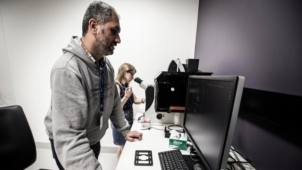
Dr Moeava Tehei at the Australian Synchrotron in Melbourne.
ABOUT THE RESEARCH
‘‘, by Elette Engels, Nan Li, Jeremy Davis, Jason Paino, Matthew Cameron, Andrew Dipuglia, Sarah Vogel, Michael Valceski, Abass Khochaiche, Alice O’Keefe, Micah Barnes, Ashley Cullen, Andrew Stevenson, Susanna Guatelli, Anatoly Rosenfeld, Michael Lerch, Stéphanie Corde and Moeava Tehei, is published in Scientific Reports.
The research was supported by funding from the Australian Government Research Training Program Scholarship, and the Australian ³Ô¹ÏÍøÕ¾ Health & Medical Research Council.


